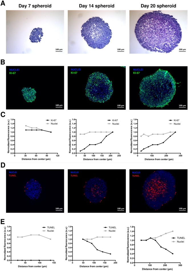
Fig 2.
(A) H&E staining of spheroid sections. (B) Determination of proliferative cells on spheroid sections by immunohistochemistry using the proliferation marker Ki-67 (green). Blue represents nuclei staining (C) Analysis of stained sections was used to compare the distribution of nuclei and proliferative cells throughout each section. (D) TUNEL assay (red) on spheroid sections to detect apoptotic cells. Blue represents nuclei staining (E) Analysis of stained sections was used to compare the distribution of nuclei and apoptotic cells throughout each section.

