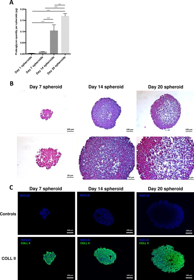
Fig 4.
(A) GAG quantification in spheroids using sulfated glycosaminoglycan assay kit. (B) Alcian blue staining of Day 7, Day 14 and Day 20 spheroid sections. (C) Immunofluorescence of type-2 collagen (green) performed on Day 7, Day 14 and Day 20 spheroid sections. Blue represents nuclei staining. Controls were obtained by omitting primary antibody but using comparative illumination parameters.

