
Figure 1. PRAME Expression in Primary Uveal Melanoma (UM) and Correlation With Survival.
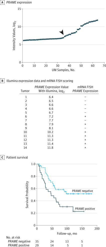
A, PRAME expression determined using 2 different probes in 64 cases of UM using an Illumina HT-12v4 microarray. Using probe ILMN_1700031, tumors are dichotomized into negative and positive. The samples on the right of the arrow are categorized as positive PRAME expression and the samples on the left of the arrow are categorized as negative PRAME expression. B, Illumina expression data and messenger RNA (mRNA) fluorescence in situ hybridization (FISH) scoring for 14 primary UMs demonstrating specificity and sensitivity of the PRAME probe sets. The κ value for mRNA FISH between both observers was 0.857. Plus sign indicates positive expression; minus sign, negative expression. C, Survival curve of patients with negative and positive PRAME-expressing primary UM.