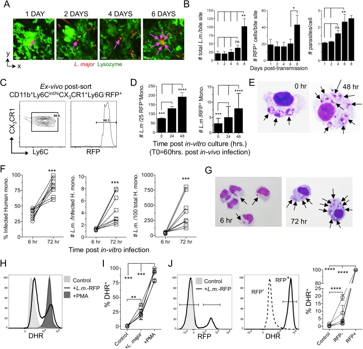
Fig 2. L. major undergoes delayed proliferation until transitioning into monocytes that are permissive for parasite replication.
(A and B) LysM-gfp mice were exposed to the bites of L. major-RFP infected sand flies and individual inoculation sites were sequentially imaged employing 2P-IVM. (A) Representative images from the same bite site of a putative single infected cell from 1 to 6 days post-bite. (B) Enumeration of the number of parasites and infected cells (parasites are assumed to be intracellular) per individual bite site employing slice-by-slice visual analysis of the z-stack to determine parasite and infected cell number. Data are the Mean +/- SEM, n = 7 bite sites. (C-E) CX3CR1-gfp mice were needle inoculated with 2x10⁵ L. major-RFP parasites in the ear. Sixty hours post-infection CD11b+Ly6Cint/hiCX3CR1+Ly6G-RFP+ cells were cell-sorted and placed in in-vitro culture. (C) Post-sort analysis of the RFP+ sorted population. (D) Quantitative cytospin analysis of the total number of amastigotes per 25 RFP+ monocytes (left panel; 100 total monocytes per time point, a denominator of 25 was chosen arbitrarily) or the number of amastigotes per individual monocyte (right panel; n = 100 (d0); 123 (24h) or 179 (48 h), total monocytes). Data are the mean +/- SD. Data are from one experiment representative of 3 similar experiments. (E) Representative cytospin images at 0h and 48h of RFP+ murine monocytes. (F-J) In-vitro L. major infection of human elutriated monocytes. (F) Percent infected, number of parasites per each infected, and total number of L. major per 100 total, monocytes. Each data set is an individual experiment employing an individual donor (n = 8), employing ≥108 infected monocytes per time point per donor. (G) Representative cytospin images. (H-J) Respiratory burst in human monocytes employing DHR. (H) Representative histogram of DHR expression by total elutriated monocytes under the indicated conditions. (I) % DHR+ monocytes under the indicated conditions. (J) Monocytes were identified as uninfected or infected based upon RFP expression (left panel) and the frequency of DHR+ cells (middle panel) was determined (right panel). In (H-J) data are from 5 experiments employing 5 individual donors.

