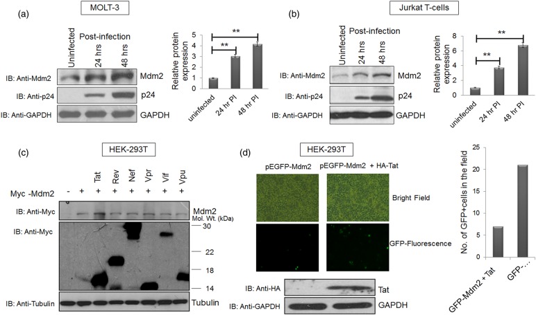
Figure 1. HIV-1 infection increases Mdm2 levels in a Tat-dependent manner.
(a) MOLT-3 cells were infected with VSVG-pseudotyped HIV-1 at an MOI (multiplicity of infection) of 1 for the indicated time periods. Cell lysates were prepared at 24 and 48 h post-infection, and western blot analysis was performed to assess total Mdm2 levels. GAPDH was used as a loading control. (b) Jurkat E6.1 cells were infected with VSVG-pseudotyped HIV-1 at MOI of 1 for 24 and 48 h. Cell lysates were prepared as described previously and western blot analysis of total Mdm2 was performed. GAPDH was used as a loading control. Bar diagrams represent the protein quantitation of the respective western blots using Image J software. (c) HEK-293T cells were co-transfected with p-CMV-Myc3-Mdm2 (2 µg) and one of the six viral regulatory/accessory gene constructs (2 µg each). Cells were collected 36 h post-transfection and subjected to western blot analysis. The bands of the viral proteins were obtained at their respective molecular sizes (Myc-Tat∼17, Myc-Rev∼19, Myc-Nef∼27, Myc-Vpr∼15, Myc-Vif∼23 and Myc-Vpu∼16; all in kDa). Tubulin was used as a loading control. (d) pEGFP-Mdm2 (3 µg) was transfected in HEK-293T cells either alone or with HA-Tat (1 µg) and the fluorescence of pEGFP-Mdm2 was assessed in the presence or absence of Tat. Expression of Tat was assessed by western blot analysis using anti-HA antibody with GAPDH as a loading control. The results are representative of three independent experiments. P-values were calculated by a two-tailed t-test (**P < 0.01).

