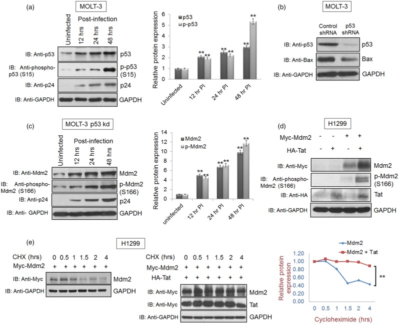
Figure 4. HIV-1 Tat-mediated Mdm2 increase is p53 independent.
(a) MOLT-3 cells were infected at an MOI of 1 with VSVG-pseudotyped HIV-1. The cells were collected at 12, 24 and 48 h post-infection and subjected to western blot analysis with anti-p53 and anti-phospho-p53 (S15) antibodies. GAPDH was used as a loading control. (b) MOLT-3 cells were transfected with shRNA scrambled (control) or p53-specific shRNA (20 µg/100 mm Petri dish) and selected using puromycin (5 µg/ml) as described in materials and methods section. The cells were collected after selection in puromycin-rich media and subjected to western blot analysis with p53 antibody. Bax was used as a positive control for the p53 knockdown experiment. GAPDH was used as a loading control. Band intensities of p53, p-p53 and GAPDH were measured by Image J software. p53/GAPDH and p-p53/GAPDH ratios were calculated for each sample. After normalisation, values of p53 and p-p53 for uninfected/control sample were assigned a value of 1 unit and compared with those of other samples in order to obtain the relative protein expression levels, as shown in the bar diagram. (c) MOLT-3 p53 kd cells were infected with VSVG-pseudotyped HIV-1 at an MOI of 1. The cells were collected at 12, 24 and 48 h post-infection and subjected to western blot analysis with anti-Mdm2 and anti-phospho-Mdm2 (S166) antibodies. GAPDH was used as a loading control. Band intensities of Mdm2, p-Mdm2 and GAPDH were measured by Image J software. Mdm2/GAPDH and p-Mdm2/GAPDH ratios were calculated for each sample. After normalisation, values of Mdm2 and p-Mdm2 for uninfected/control sample were assigned a value of 1 unit and compared with other samples in order to obtain the relative protein expression levels, as shown in the bar diagram. (d) H1299 p53-null cells were transfected with 2 µg of Myc-Mdm2 alone or in the presence of HA-Tat, and the cell lysates were collected 36 h post-transfection. Anti-Mdm2 and anti-phospho-Mdm2 (S166) antibodies were used for western blot analysis. (e) Cycloheximide chase assay was performed to assess the effect of Tat on Mdm2 stability in the absence of p53 using H1299 (p53-null) cells by treating the cells with 100 µg/ml of CHX 36 h post-transfection for the indicated time periods. The results are representative of four independent experiments. P-values were calculated by a two-tailed t-test (**P < 0.01).

