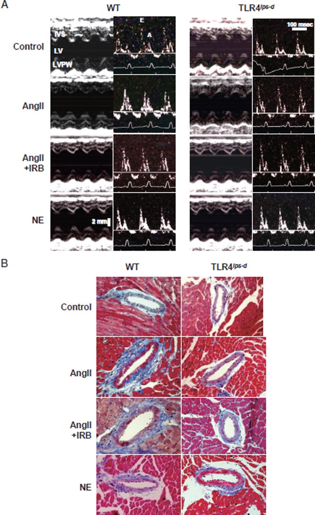
Fig. 1. Representative Echocardiography and Micrographs of Vascular Remodeling and Perivascular Fibrosis in the Intramyocardial Arteries.
WT, wild-type mice; TLR4lps-d, TLR4-deficient mice; AngII, angiotensin II; NE, norepinephrine; IRB, irbesartan.
A. Representative echocardiograms of WT and TLR4lps-d mice. AngII and NE were infused subcutaneously for two weeks using an osmotic mini-pump. The cardiac function was evaluated using transthoracic echocardiography with an ultrasound machine equipped with a 15-MHz probe under light anesthesia and sevoflurane in the WT and TLR4lps-d mice. Left panel: M-mode recording of the left ventricle. Right panel: transmittal flow assessed on Doppler echo. IVS, interventricular septum; LV, left ventricle; LVPW, left ventricular posterior wall; E, early ventricular filling wave (E wave); A, late filling wave caused by atrial contractions (A wave). B. Representative micrographs of the effects of TLR4 on vascular remodeling and perivascular fibrosis in the intramyocardial arteries. The sections were stained with Masson Trichrome staining. Bar, 50 µm.

