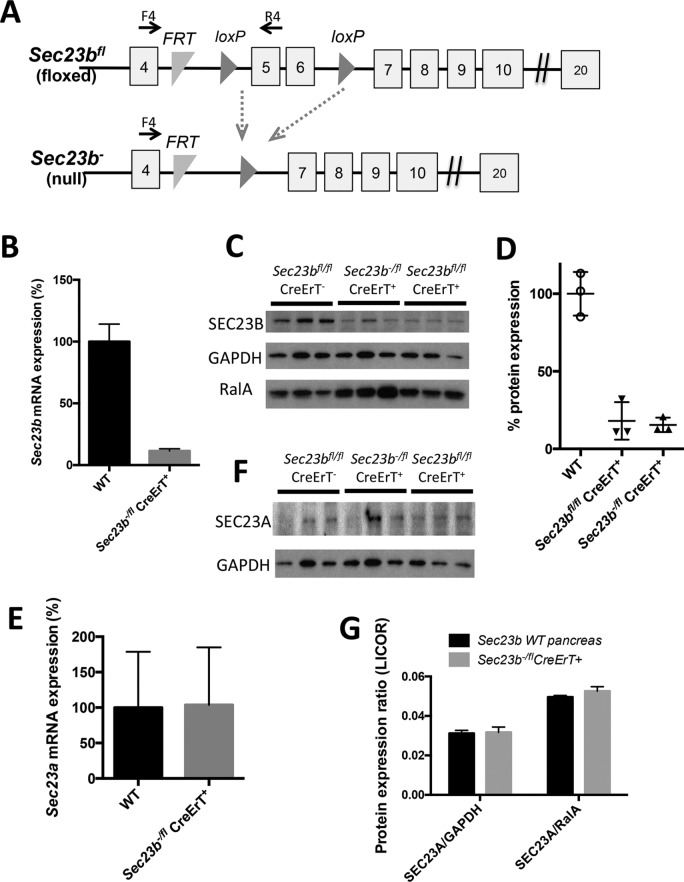
FIGURE 1:
Sec23b inactivation in pancreatic acinar cells. (A) Sec23b alleles (not drawn to scale; Khoriaty et al., 2014). Each square indicates an exon, and horizontal lines between exons indicate introns. F4 and R4 are primers used for assessing efficiency of Cre-mediated excision. (B) Sec23b excision determined by qPCR (n = 3 for each genotype) and (C) Western blot on pancreas tissues 7 d after administration of tamoxifen. (D) Quantification of the SEC23B band intensities in C relative to average of GAPDH and RalA performed using ImageJ. (E–G) Quantitation of Sec23a expression by (E) qPCR (three controls and four Sec23b−/fl CreErT+ mice), (F) chemiluminescence Western blot detection, and (G) quantitative Western blot (infrared fluorescence detection) in pancreas tissues 7 d after administration of tamoxifen (three mice per genotype).

