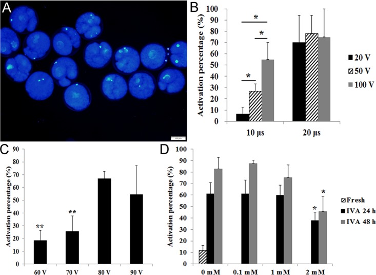
Figure 1. Effects of different stimulus conditions and concentrations of melatonin on oocyte activation.
(A) Fresh oocytes were activated by weak stimulus (70V, 10μs, 2 pulses and stained with DAPI to show female pronucleus formation. (B) Oocytes were activated by different combinations of electric pulse times (10 μs and 20 μs) with voltage intensity (20 V, 50 V and 100 V). (C) Oocytes were activated by different voltage intensity treatment (60 V, 70 V, 80 V and 90 V; 10 μs). (D) Oocyte aged for 24 hr or 48 hr in vitro were treated with different concentrations of melatonin and activated by weak stimulus set 70 V, 10 μs, 2 pulses. All graphs show mean ± s.e.m. Abbreviations used in this and all subsequent figures: IVA, in vitro aging. Independent replicates were conducted with a minimum of 25 oocytes/replicate, at least 3 stable replicates were obtained. *P<0.05, **P <0.01. Bar = 100 μm.

