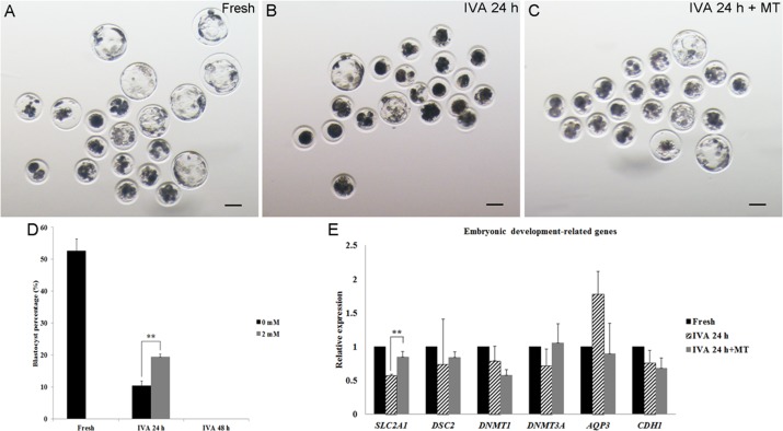
Figure 3. The development of embryos from fresh, aged and melatonin treated oocytes after parthenogenetic activation.
(A) Fresh oocytes. (B) Oocytes aged for 24 hr in vitro. (C) IVA 24 hr oocytes treated with 2mM melatonin. (D) Blastocyst formation from fresh oocytes, IVA 24 hr or 48 hr oocytes and oocytes treated with melatonin for 24 hr or 48 hr after strong stimulus (800V, 40μs, 2 pulses). (E) The expression of embryonic development-related genes (SLC2A1, DSC2, DNMT1, DNMT3A, AQP3 and CDH1) in parthenogenetic blastocysts from fresh and aged oocytes. All graphs show mean ± s.e.m. Independent replicates were conducted with a minimum of 25 embryos/replicate, at least 3 stable replicates were obtained. **P <0.01. Bar = 100 μm.

