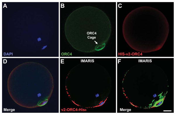
Figure 2. Incorporation of v2-ORC4-HIS6 into the ORC4 Cage.
Purified v2-ORC4-His6 protein was injected into MII oocytes prior to activation. The oocytes were developed to G1 with the second polar body, then fixed and immunostained for ORC4 (green) and for the Histidine tag (red). The figure show confocal sections of one oocytes with filters for DAPI (A), ORC4 (B), His-tag (C), and merged (D). IMARIS software was used to identify where the v2-ORC4-His6 was localized (E and F). Bar = 10 μm.

