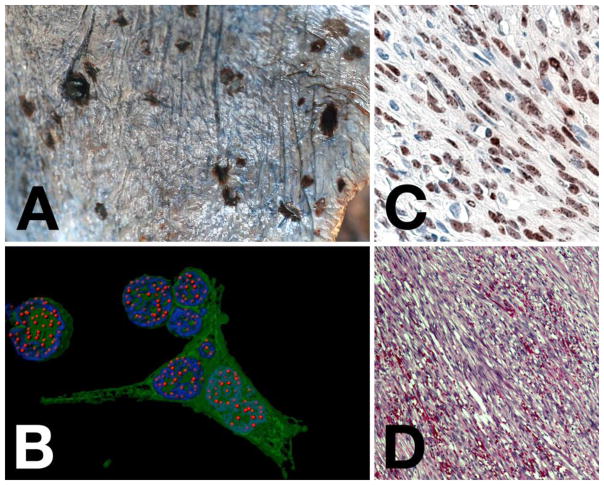
Figure 1. KS pathology and histology.
Panel A shows an image of gross morphology of disseminated KS on the surface of the lung. Note the single, raised, nodular lesion in the upper left, as compared to the flat lesions. Panel B shows a computer-enhanced image of immunofluorescence in a KSHV recombinant virus that also expressed green fluorescent protein (gfp) in a PEL cell line. LANA staining is in red, nuclear DNA staining in blue and gfp (to indicate infected cells) in green. This analysis clearly shows the presence of discrete “LANA dots”, each indicating a place where the viral genome is tethered to the host chromosome. Panel C shows an image of LANA staining of a KS lesion by immunohistochemistry (brown) with hematoxilin counterstain (blue). Note all LANA staining is nuclear and the appearance of darker spots or dots within the nuclear staining. Panel D, shows an H&E stain of a KS lesion. Note the spindle shape nature of the cells, which are of endothelial cell lineage. Slit-like spaces in between the cells contain extravasated red blood cells.

