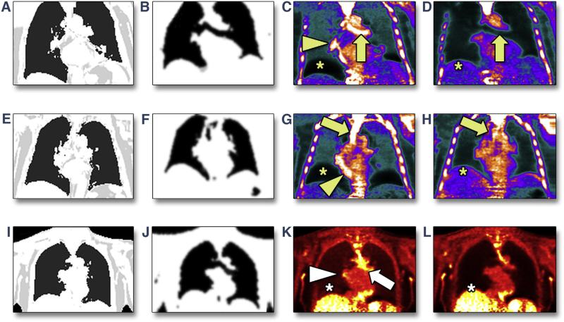
FIGURE 2. Resolution of Positron Emission Tomographic/Magnetic Resonance Artifacts Using the Novel Free-Breathing Radial Gradient-Recalled Echo Attenuation Correction Approach.
In 2 patients undergoing 18F-sodium fluoride positron emission tomographic (PET) imaging (A to D, E to H) and 1 undergoing 18F-fluorodeoxyglucose PET imaging (I–L), attenuation maps were based either on standard breath-held gradient-recalled echo (GRE) (A, E, I) or free-breathing radial GRE (B, F, J). Artifacts at the liver-lung and heart-lung boundaries and in the bronchus present in PET images reconstructed with breath-held GRE magnetic resonance attenuation correction (MRAC) (C, G, K) are removed by using the novel free-breathing radial GRE approach for MRAC (D, H, L).

