Fig. 5.
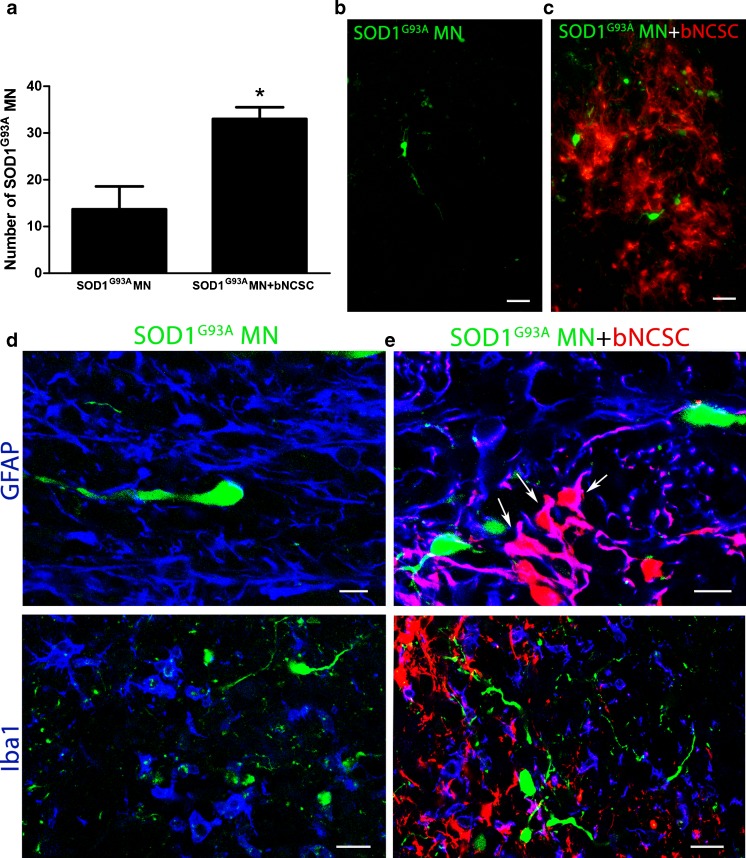
Increased survival of SOD1G93A MN after co-implantation with bNCSC to the spinal cord of mice. One week after implantation, SOD1G93A MN co-implanted with bNCSC showed significantly increased survival compared to SOD1G93A MN implanted alone. Asterisks indicate the level of statistical significance by two-tailed Student’s t-test (* p < 0.05). Data shown is in mean ± SEM of three animals per condition (a). SOD1G93A MNs when implanted alone are located as single cells in the spinal cord parenchyma (b). When implanted together with bNCSCs, SOD1G93A MNs can be found both spread out as single cells as well as in close proximity to bNCSCs (c). The injection site showed increased immunoreactivity for GFAP and Iba1 following implantation of either SOD1G93A MN alone (d) or co-implantation with bNCSCs (e). Arrows indicate bNCSCs differentiated to GFAP positive cells (e). Scale bars: b,c 25 μm; d,e 10 μm for GFAP and 20 μm for Iba1

