Figure 6.

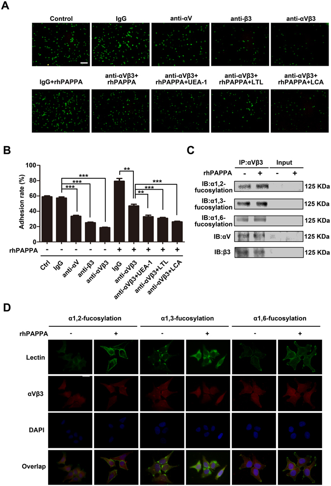
rhPAPPA promotes endometrium receptivity by up-regulating the specific fucosylation of integrin αVβ3. (A) Adhesion of JAR cells to Ishikawa cells pre-treated without (a) untreated control, or with (b) IgG, (c) anti-αV, (d) anti-β3, (e) anti-αVβ3, (f) rhPAPPA + IgG, (g) rhPAPPA + anti-αVβ3, (h) rhPAPPA + anti-αVβ3 + UEA-1, (i) rhPAPPA + anti-αVβ3 + LTL, (j) rhPAPPA + anti-αVβ3 + LCA. (B) Adhesion rate of JAR cells to pre-treated Ishikawa as indicated is shown in the histogram. (C) Integrin αVβ3 was immunoprecipitated from the whole-protein lysate of untreated control and rhPAPPA-treated Ishikawa cells. The specific N-fucosylation of αVβ3 was detected by Western blotting. αV and β3 were detected to show the loading protein amount. Input showed the efficiency of immunoprecipitation. (D) Immunofluorescence and Lectin staining detected the expression and cellular localization of the specific fucosylation and αVβ3 after rhPAPPA treatment. Green, α1,2-, α1,3-, or α1,6-fucosylation; red, αVβ3; yellow (overlay), co-staining of α1,2-, α1,3-, or α1,6-fucosylation with αVβ3. DAPI (blue) was used for nuclear staining. The bar represents 50 μm. **p < 0.01, ***p < 0.001. The data were presented as the means ± SEM of three independent experiments.
