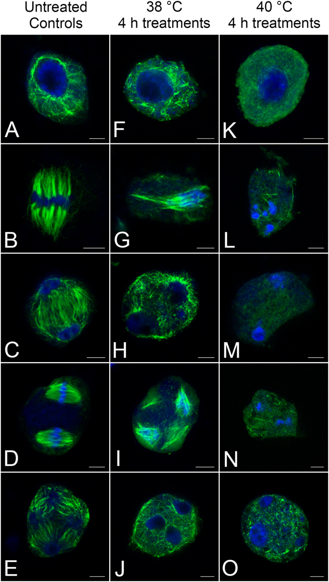
Figure 6.

Defects in microtubule distribution and chromosome behaviours induced by high temperature treatments. (A–E) Microtubular distributions and chromosome behaviours under normal conditions. (A) The network of microtubules in prophase I. (B) Connections between chromosomes and microtubules in a metaphase I dipolar spindle. (C) Phragmoplast microtubules located between two daughter nuclei in telophase I. (D) Two spindles with parallel arrangement in metaphase II. (E) Nuclear-based RMSs and four separated daughter nuclei in telophase II. (F–J) Defects in microtubular arrangement and abnormal chromosome behaviours after treatment at 38 °C for 4 h. (F) Microtubules become fragments in prophase I. (G) Metaphase I spindle damage, resulting in unrestrained chromosomes in the cytoplasm. (H) High temperature treatment resulting in destroyed phragmoplast with a micronucleus. (I) Damaged spindles in metaphase II. (J) Failure of RMS formation due to heat stress. (K–O) Damages to the microtubular arrangement and chromosome behaviours after treatment at 40 °C for 4 h. (K) A cell in prophase I shows almost no microtubules. (L) A few microtubular fragments remaining in the cytoplasm of a cell in metaphase I. (M) Completely depolymerised microtubules in telophase I. (N) No visible spindles and just a few microtubular fragments in metaphase II, resulting in excessive condensation of chromosomes. (O) Depolymerised microtubules causing fusion of nuclei and formation of micronuclei in telophase II. Bars = 5 μm.
