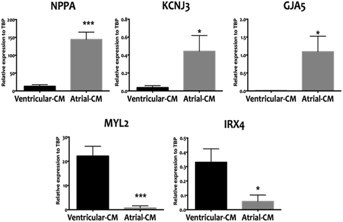
Figure 1.

qRT-PCR-based expression analyses directly comparing the atrial differentiation protocol that employed RA (atrial-CM, grey bars) and ventricular differentiation protocol (ventricular-CM, black bars). Cell populations were analyzed at T20 of the differentiation protocol. The RA protocol was able to generate cells that are enriched in atrial specific markers (NPPA, KCNJ3, and GJA5) and lacked ventricular markers (MYL, IRX4). Values are shown relative the housekeeping gene TBP. Error bars represent standard deviation of the mean from the values of independent experiments (N ≥ 4); *P ≤ 0.05, **P ≤ 0.01, ***P ≤ 0.001.
