Figure 6.

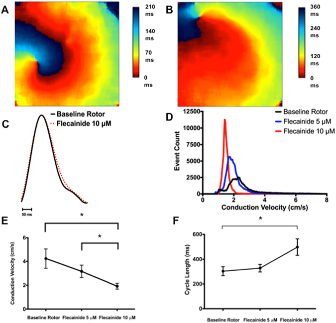
The effects of flecainide on rotor dynamics, APD, CV and CL in atrial-CM sheets. (A) and (B) Show typical activation maps of an atrial-CM sheet in the absence (A) and presence (B) of 10 μM flecainide. Notice the spreading of the activation times when flecainide is present indicating a slowing of the CV. C) shows AP traces, average over the entire sheet) in the absence and presence of 10 μM flecainide. Flecainide did not cause APD prolongation. (D) Presents a histogram of CVs measured at all points in a CM sheet prior to and after the administration of 5 or 10 μM flecainide. Flecainide shifted the histogram distributions of CVs to lower values as a function of drug concentration. (E) Summarizes the CV in 7atrial-CM sheets as a function of flecainide. (F) Shows a summary of the CL for the same sheets and conditions shown Panel (E). APD = action potential duration, CV = conduction velocity, CL = cycle length. *p < 0.05.
