Figure 6.

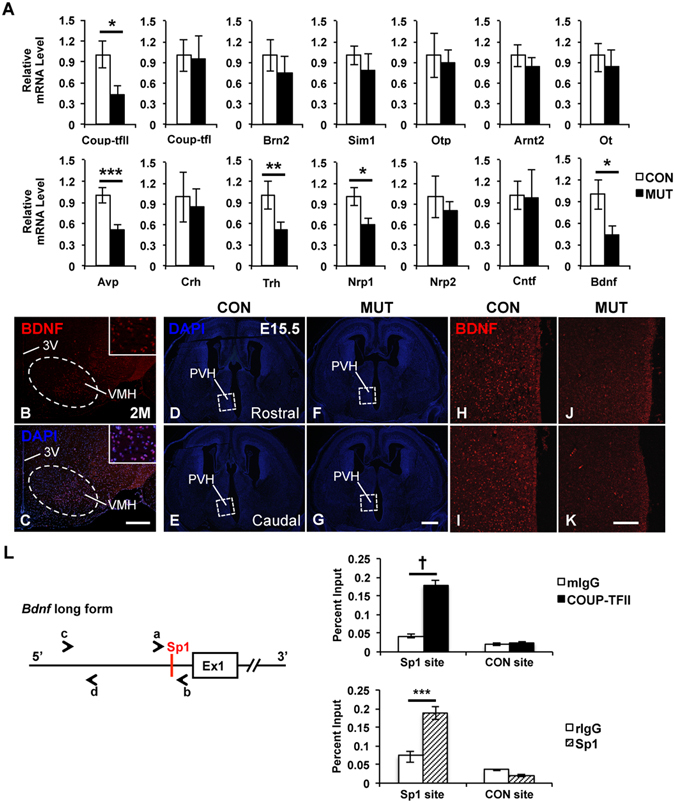
Expression of Bdnf and Nrp1 is reduced in the COUP-TFII mutant embryo, and Bdnf is a downstream target of COUP-TFII. (A) Real-time quantitative PCR data with samples from the ventral forebrain of the control (n = 4) and mutant (n = 3) embryos at E14.5. The expression of COUP-TFII, Avp, Bdnf, Nrp1 and Trh transcripts is significantly reduced in the mutant. A BDNF-specific antibody detects the expression of BDNF in the cytoplasm of neurons at the ventro-lateral region of the mouse VMH nucleus at 2 M (B,C). DAPI staining images of coronal sections with the PVH region in the control (D,E) and the mutant (F,G) at E15.5. Compared with the control (H,I), the expression of BDNF protein is noticeably reduced in the prospected mutant hypothalamus at E15.5 (J–K). L, An evolutionarily conserved Sp1 binding site is identified at the promoter region of the long form of the Bdnf gene. Primers a/b and c/d were used to amplify the Sp1 locus and a 2 kb up-stream non-Sp1 control locus, respectively. In chromatin immunoprecipitation assays, the binding of both COUP-TFII and Sp1 protein is enriched at the conserved Sp1 site but not at the negative control site. 3 V, third ventricle; PVH, paraventricular nucleus of hypothalamus; VMH, ventromedial nucleus of hypothalamus. Three independent assays were performed in each real-time quantitative PCR experiment. The data indicate the mean ± SD. Student’s t-test, *P < 0.05; **P < 0.01; ***P < 0.005; †P < 0.001. Scale bar, (B,C) 300 μm; (D–G) 500 μm; (H–K) 100 μm.
