


Figure 1.
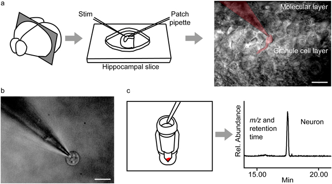
Patch clamp-assisted single neuron lipidomics. (a) Mouse brain slices are prepared using a vibratome and transferred to the perfusion chamber of a standard electrophysiology setup. A target neuron is visually identified and patched in the cell-attached configuration using a standard manipulator. A patched granule cell of the dentate gyrus and the attached pipette are shown in pseudocolor. (b) The neuronal soma is carefully pulled away from the slice while maintaining the giga-ohm seal. Visual inspection is used to confirm that the neuron is free of extraneous contaminating material such as glial cells; calibration bar: 10 μm (c) The patched neuron is aspirated into the pipette tip and transferred to a sterile vial kept in dry ice. Stimulated cells are collected from the slice within 3 min of stimulation. Samples are subjected to single-phase isopropanol extraction and lipids are analyzed by nanoflow liquid chromatography/high-resolution time-of-flight mass spectrometry (nLC-MS).