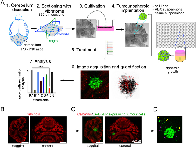
Figure 1.

The organotypic cerebellar slice–tumour co-culture. (A) Workflow for OCSC generation and tumour spheroid implantation. (1) Decapitation of mouse pup(s) at PND 8–10 and isolation of cerebellum. (2) Sectioning of cerebellum under physiological conditions using a vibratome to generate 350 µm thick sagittal or coronal sections. (3) Culture of cerebellum sections on membrane inserts for 15 days in vitro (DIV). (4) Implantation of one LifeAct enhanced GFP (LA-EGFP) expressing tumour spheroid per slice. (5) Further incubation of the co-culture in vitro without or with treatment. (6) Fixation and immunofluorescence for microscopy analysis. (7) Quantification and data analysis. (B) Sagittal and coronal sections of cerebella, stained with anti-Calbindin (red) to visualise Purkinje cells. (C) Implantation of LA-EGFP expressing DAOY cells spheroid on the slices. (D) Dissemination of tumour cells (green) in the cerebellar slices after 5 days post initiation of culture.
