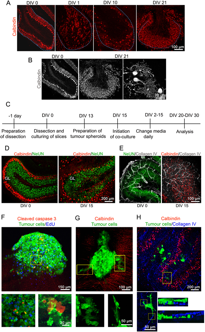
Figure 2.

Morphological assessments of the cerebellar slices and co-culture in vitro. (A) Changes in the Purkinje cell monolayer upon in vitro culturing of the slices was monitored at DIV 0, DIV 1, DIV 10 and DIV 21 with anti-calbindin antibody (red) using confocal microscopy. (B) Grey scale images of cerebellar slices at DIV 0 and after being cultured for DIV 21. (C) Timeline of the experimental setup for five-day co-culture experiment. DIV 15 was selected as the time point suitable for the implantation of tumour spheroid. (D) The cerebellar cortex layers visualised using confocal microscopy at DIV 0 and DIV 15 for comparison with anti-Calbindin (red, Purkinje cell layer) and anti-NeuN (green, granular layer) antibodies. (E) Visualisation of the vasculature using anti-collagen IV (grey) at DIV 0 and DIV 15. (F) Determination of apoptosis and proliferation after five-day co-culture using anti-cleaved caspase 3 (red) and Click-iT EdU (blue), respectively. (G) Monitoring the tumour cells infiltrating the cerebellar slices. (H) Visualization of the vasculature (blue) in the presence of tumour cells. Lower panels: zoomed versions of boxed insets in F, G and H respectively. In H, EGFP and Collagen IV fluorescence was volumised.
