
Figure 2.
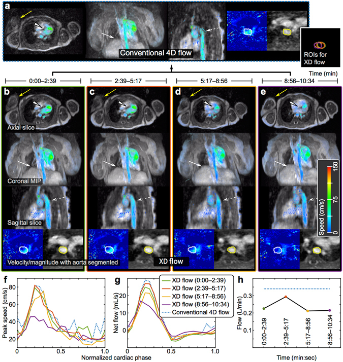
XD flow reconstruction of a 3-day-old female highlighting enhanced motion robustness. (a) Conventional 4D flow reconstruction for the 10:34 min acquisition. (b–e) The same dataset reconstructed into 4 shorter temporal windows — each window was 2:39 min. (f) Peak speed in aorta. (g) Net flow in aorta. (h) Average flow for each temporal window compared to conventional 4D flow (dotted blue). Different reformats of a single cardiac phase are shown: axial slice, coronal 50-cm MIP, sagittal slice, and velocity/magnitude of aorta with segmentation. The non-sedated patient with ferumoxytol enhancement was observed to be moving as noted by different positions of the right arm (yellow arrow). Also, the region-of-interests (ROIs) segmented for each temporal phase of XD flow are combined to emphasize the movement of the aorta (far right of (a)). With motion corruption, the flow in the aorta is noisy with unrealizable flow vectors in the conventional 4D flow, but the flow is recovered in the XD flow reconstruction (dashed white). Similarly, the myocardial border (white triangle) and diaphragm (white arrow) are better depicted in the XD flow reconstruction. The blood flow measured from XD flow varies over time; this effect is ignored in conventional 4D flow.