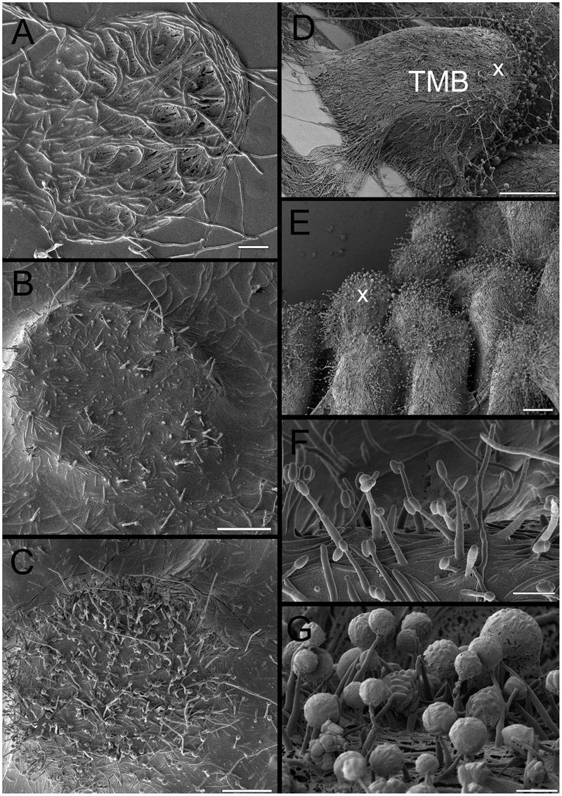
FIGURE 7.

Overview of cryo-SEM image of developing TMB. (A) Trapping mycelia in a 180 μm diameter well after 2 days. (B) Growth out of the well after 3 days with limited dehydration prior to imaging, emphasizing the high degree of hydration. (C) As (B), but at a slightly more advanced stage and with more removal of water revealing hyphae and some phialides. (D) Mature TMB after 8 days showing most phialides at the apex (x). (E) As (D) but showing multiple TMB. (F) Phialides after 3 days. (G) Close-up of phialides at apex of the TMB after 8 days. Note the large spherical clusters containing large numbers of conidia. Scale bar indicates 40 μm for (A–C), 140 μm for (D), 60 μm for (E), and 5 μm for (F,G).
