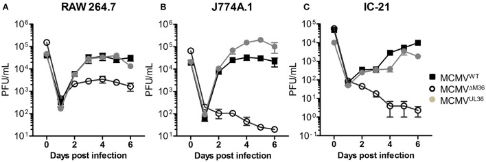
Figure 5.
UL36 protein rescues growth of ΔM36 MCMV in macrophages. Growth of MCMVWT, ΔM36.MCMV and MCMVUL36 was compared in (A) RAW 264.7, (B) J774A.1, and (C) IC-21 macrophage cell lines. RAW 264.7 cells were infected at an MOI of 2.5 while J774.A and IC-21 were infected at 1 MOI. The infectious virus in supernatants was quantified plaque assay on C57BL/6 MEF cells. Values represent means of three biological replicates, while error bars indicate SEM.

