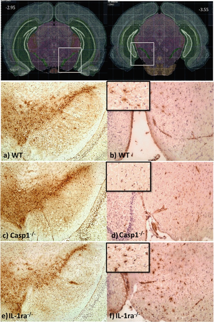
Fig. 3.
Immunostaining of TH-positive neurons and CD68-positive microglia cells. Frozen sections of mouse brains were stained with anti-TH and anti-CD68 antibody at 15 months. Positive TH immunostaining of a wt, c casp1−/−, and e IL-1ra−/−, and positive CD68-immunostaining of b wt, d casp1−/−, and f IL-1ra−/− mice

