
Figure 3. An elevated endogenous NMDAR tone in RVH rats tonically inhibits I A .
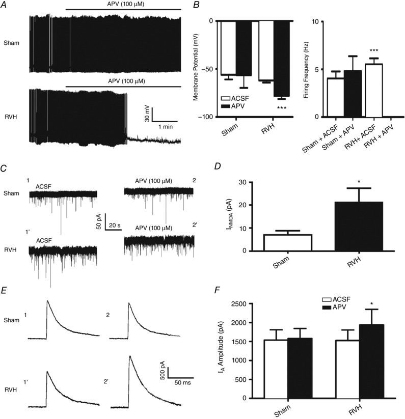
A, representative samples from MNCs from a sham and RVH rat showing that blockade of NMDAR (APV 100 μm) strongly hyperpolarized and inhibited ongoing firing activity in the RVH but not in the sham rat. B, summary data of mean membrane potential (V m) and firing frequency in MNCs from sham and RVH rats, n = 5 and 8, respectively) before and after APV application. C, representative samples from MNCs from a sham and RVH rat showing that blockade of NMDARs (APV, 100 μm) induced an outward shift in the holding current (I NMDA). The representative segments shown were taken just before APV application (ACSF) and 8.5 min in APV. Note the larger I NMDA observed in the MNC from the RVH rat. D, summary data of mean I NMDA in MNCs from sham and RVH rats (n = 8 and 13, respectively). E, representative examples of I A (command step to −20 mV) before and during APV (100 μm) in the same sham and RVH rat as in A. Time points 1 and 2 correspond to the same ones as shown in C. F, summary data of mean I A amplitude evoked before and during NMDAR blockade (APV) in MNCs from sham and RVH rat (n = 8 and 13, respectively). * P < 0.05 and *** P < 0.0001 vs. respective control.