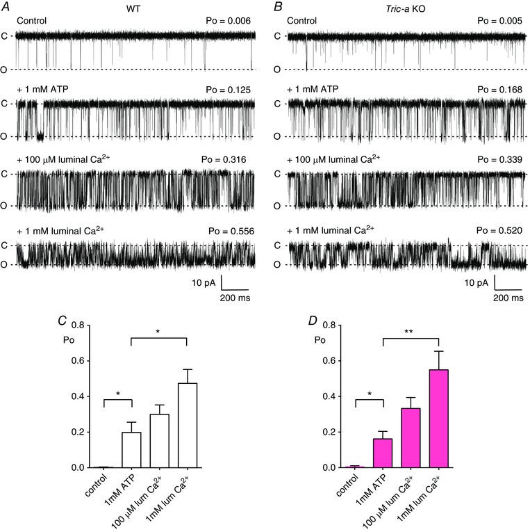
Figure 1. The effects of cytosolic ATP and luminal Ca2+ on RyR1 channels from Tric‐a KO mice with K+ as the permeant ion.

Representative single‐channel recordings of RyR1 from WT (A) and Tric‐a KO (B) mice under control conditions (10 μm cytosolic and luminal Ca2+), after subsequent addition of 1 mm cytosolic ATP (second traces) and subsequent increasing concentrations of luminal Ca2+ (100 μm and 1 mm as indicated). The bar charts below illustrate the mean data for RyR1 channels from WT (white) (C) and Tric‐a KO (pink) (D) skeletal muscle under control conditions, in the presence of ATP and increasing concentrations of luminal Ca2+. Values are the mean ± SEM (n = 6–10; * P < 0.05, ** P < 0.01). The holding potential was −30 mV. O and C indicate the open and closed channel levels, respectively.
