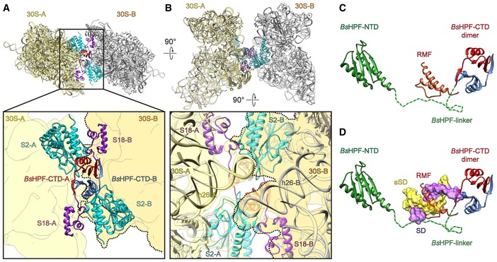
Figure 4. Dimerization interface of the Bs70S‐30S subcomplex.

-
A, BDistinct views of the dimer interface between 30S‐A (yellow) with BsHPF‐CTD‐A (red) and 30S‐B (gray, darker yellow with dashed line in zoomed panel) with BsHPF‐CTD‐B (blue). Ribosomal proteins S2 (cyan), S18 (purple), and 16S rRNA are shown only, and the surface outline of the 30S subunit is included schematically for reference.
-
C, DBinding site of BsHPF‐NTD (green) and dimeric BsHPF‐CTD (red, blue) relative to (C) RMF (orange; Polikanov et al, 2012) and (D) SD–anti‐SD helix (yellow‐purple surface; Sohmen et al, 2015). The dashed line indicates the linker and is shown only to illustrate that the 34 amino acids are more than sufficient to connect the NTD and CTD; however, no density for the linker was observed, suggesting it does not adopt a defined conformation on the ribosome.
