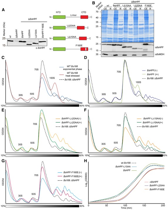
Figure 5. Monitoring 100S formation in vivo for BsHPF variants.

-
AWestern blot using antibodies raised against BsHPF to assess the levels of BsHPF in cell extracts of wild‐type Bs168 (wt), ∆BsHPF, and ∆BsHPF strains expressing either wild‐type BsHPF or BsHPF‐L∆20AA, BsHPF‐L∆10AA, BsHPF‐F160E variants.
-
BCoomassie (above) and Western blot of cell extracts (CE) and ribosome pelleted fractions (R) of the wild‐type Bs168 (wt) strain or the ∆BsHPF strains expressing either wild‐type BsHPF, BsHPF‐L∆10AA, BsHPF‐L∆20AA, and BsHPF‐F160E.
-
CSucrose gradient profiles of cell extracts from the wild‐type Bs168 (wt) strain in exponential phase (blue) or heat stressed (red), compared with the extract from the Bs168 ∆BsHPF strain (dashed line).
-
D–GSucrose gradient profiles of cell extracts from the (D) Bs168 ∆BsHPF amyE::BsHPF strain, (E) Bs168 ∆BsHPF amyE::BsHPF‐L∆20AA strain, (F) Bs168 ∆BsHPF amyE::BsHPF‐L∆10AA strain, and (G) Bs168 ∆BsHPF amyE::BsHPF‐F160E strain in the absence (I−) or presence (I+) of IPTG. The dashed line of the Bs168 ∆BsHPF strain from (C) is shown for reference.
-
HGrowth curves illustrating the recovery from stationary phase of the wild‐type Bs168 (wt), ∆BsHPF, and ∆BsHPF strains expressing either wild‐type BsHPF or BsHPF‐L∆20AA, BsHPF‐L∆10AA, BsHPF‐F160E variants.
Source data are available online for this figure.
