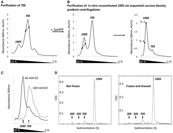
Figure 1. Purification and characterization of 100S ribosome dimers.

- Sedimentation profile performed to separate 70S ribosomes from 100S particles present in the cell.
- Sedimentation profiles were carried out sequentially in order to separate 100S from 70S particles. SaHPF was added to fractions containing 70S particles (intercept on panel A). Fractions containing 100S dimers (intercept on left panel) were further purified on a similar gradient (right panel). Fractions 4 and 5 were used to assess stability and to perform cryo‐EM analysis.
- 100S dimers are stable at low ionic strength. Sucrose gradient performed under similar conditions to that shown in panel (B).
- 100S dimers do not suffer from freezing. The sedimentation profiles from analytical ultracentrifugation experiments are shown for a sample that was either kept at 4°C prior to analysis (left), or frozen at −80°C and thawed at 4°C (right).
