
Figure EV3. Effect of mutations in XPF‐ERCC1 on ICL repair in Xenopus egg extract.
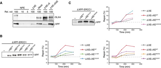
- Mock‐depleted, XPF‐ERCC1‐depleted (ΔXE), and XPF‐ERCC1‐depleted NPE complemented with SLX4 (ΔXE+S) or XPF‐ERCC1 and SLX4 (ΔXE+SXE) were analyzed by Western blot using α‐XPF or α‐SLX4 antibodies. A dilution series of undepleted NPE was loaded on the same blot to determine the degree of depletion. A relative volume of 100 corresponds to 0.2 μl of NPE.
- Replicates of Fig 2B. XPF‐ERCC1‐depleted (ΔXE) and XPF‐ERCC1‐depleted extracts complemented with wild‐type (XEWT) or indicated mutant XPF‐ERCC1 (XEMUT) were analyzed by Western blot using α‐XPF antibodies (left panel). These extracts were used to replicate pICL. Replication intermediates were isolated and digested with HincII, or HincII and SapI, and separated on an agarose gel. Repair efficiency was calculated and plotted (right panels).
- As in (B) but analyzing different mutant complexes. Note: repair levels can differ per batch of individually prepared extract or per depletion experiment and can only be compared within an experiment.