
Figure 2.
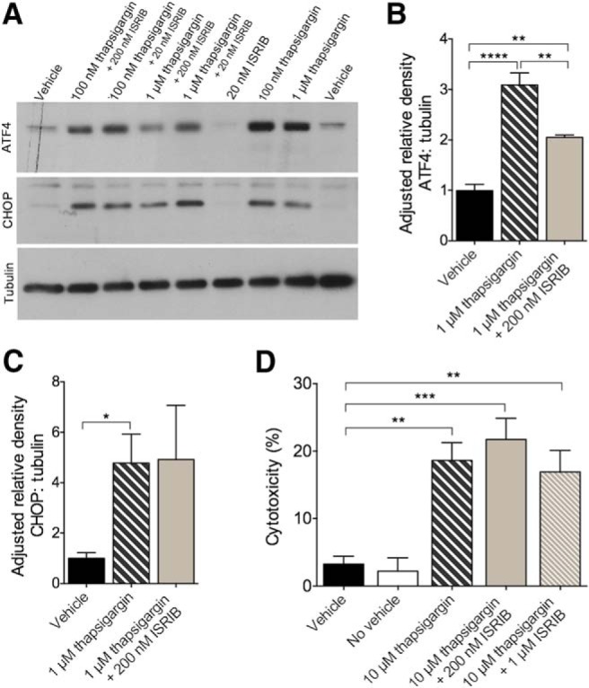
Thapsigargin induced ER stress and target engagement in vitro. A–D, ER stress-induced ATF4 translation, but not CHOP activation or cytotoxicity, is reduced by ISRIB in rat PCNs. A, Representative immunoblots of primary cortical cell lysates derived from E17 Sprague Dawley rats probed using antibodies directed against ATF4, CHOP, and tubulin. B, Quantification of ATF4 levels normalized to tubulin. ATF4 is increased in cells treated with 1 μM thapsigargin or 1 μM thapsigargin + 200 nM ISRIB compared to vehicle control. Cells treated with 1 μM thapsigargin have more ATF4 compared to cells treated with 1 μM thapsigargin + 200 nM ISRIB. Vehicle, n = 6; 1 μM thapsigargin, n = 5; 1 μM thapsigargin + 200 nM ISRIB, n = 4. C, Quantification of CHOP levels normalized to tubulin. CHOP is increased in cells treated with 1 μM thapsigargin compared to vehicle control. Vehicle, n = 4; 1 μM thapsigargin, n = 3; 1 μM thapsigargin + 200 nM ISRIB, n = 3. D, Quantification of cytotoxicity by LDH. Cells treated with 10 μM thapsigargin, 10 μM thapsigargin + 200 nM ISRIB, or 10 μM thapsigargin + 1 μM ISRIB have higher percentages of cytotoxicity compared to vehicle control. Vehicle, n = 6; no vehicle, n = 6; 10 μM thapsigargin, n = 6; 10 μM thapsigargin + 200 nM ISRIB, n = 6; 10 μM thapsigargin + 1 μM ISRIB, n = 6. Error bars indicate SEM; *p < 0.05, **p < 0.01, ***p < 0.001, ****p < 0.0001.