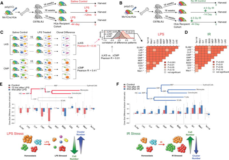
Figure 4. Hematopoietic Cell Autonomy Is Persistent upon Stress.

(A) The experimental design to study hematopoietic response upon LPS-mediated inflammatory stress. A HUe recipient cohort was generated by transplanting aliquots of the same mixture of randomly labeled HSPCs into 15 lethally irradiated C57BL/6J recipients. After 16 weeks of reconstitution, the cohort was divided into three sub-cohorts. One sub-cohort received LPS injection 12 hr priorto analysis (12hrs), another sub-cohort at 44 days prior(44 day), and a third sub-cohort received PBS treatment (control). All mice, including the control group, were sacrificed on the same day and assayed by flow cytometry.
(B) The experimental design to study the effect of genotoxic stress on hematopoietic clonal dynamics. An independent HUe recipient cohort was generated as in (A). After16 weeks of hematopoietic reconstitution, the cohort was divided into three sub-cohorts:a control group that received no irradiation, one group received 4.5 Gy irradiation 14 days prior to data collection (14 day), and another group received 4.5 Gy irradiation at 44 days prior to data collection (44 day). All mice, including the control group, were harvested for data collection on the same day.
(C) To test for consistency of clonal response to LPS treatment among cohort recipients, we performed pairwise comparisons of the fluorescent clonal pattern for each hematopoietic compartment (e.g., SLAM, LKS, CLP, etc.) and across each LPS-stressed and control animal. For all pairwise comparisons,the correlation of the LPS-associated clonal changes was significantly higher within a compartment than between compartments. In other words, the clonal response to the LPS insult was distinct among individual hematopoietic cell types, but was highly consistent across multiple HUe recipients (*p < 10−7 significance when comparing a given cell type with all other cell types).
(D) Consistency of the clonal response to irradiation treatment was assessed in the same way as in (C). For the vast majority of pairwise comparisons, the clonal changes within a cell compartment showed significantly higher correlation than between cell compartments. Each cell type was significantly distinct when compared against all other cell types (*p < 10−7) but consistent across multiple recipients.
(E) LPS treatment led to reduction in output of existing clones. Illustration of the hematopoietic clonal response to LPS inflammatory stress at 12 hr and day 44 in comparison to saline-treated controls at the stem, progenitor, and mature stages. To quantify the effect of LPS treatment, we performed pairwise comparison of mice from LPS-treated and control groups, detecting parts of the fluorescent spectra showing statistically significant differences in cell density. The barplot shows average change of cell numbers (measured as a fraction of the total number of cells measured) within such regions (whiskers show 95% confidence interval). The negative values correspond to decrease in the cell counts relative to control group. The analysis shows that at 12 hr following LPS treatment, the majority of existing clones were significantly reduced in size, accompanied by appearance of small new clones (Figure S5D). Such changes preferentially impacted the HSC-MPP-CMP-GMP branch of hematopoiesis, and were largely attenuated at 44 days post LPS treatment.
(F) Irradiation triggered expansion of existing clones. The barplots show the prevalent direction of cell density changes at 14 or 44 days following irradiation treatment. The changes were most pronounced at 44 days and showed widespread increase in the output of individual clones, significantly affecting most of the hematopoietic cell types.
See also Figure S3.
