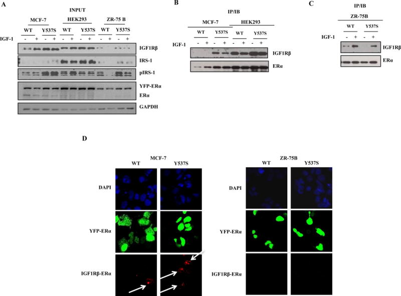
Figure 4. MCF-7 Y537S cells show the lowest anti-proliferative response to Tam through increased interaction between IGF1R/ER.

a, b, and c, Immunoblot analysis to detect expression of IGF1Rβ, pIRS-1, IRS-1 and ER in input extracts; GAPDH was used as a loading control (a). Coimmunoprecipitation assays of IGF1R/ER interactions (b and c). Duolink staining (red) demonstrated binding between IGF1R and ER in MCF-7(d) and ZR-75B cells (e). Images are representative of three different experiments.
