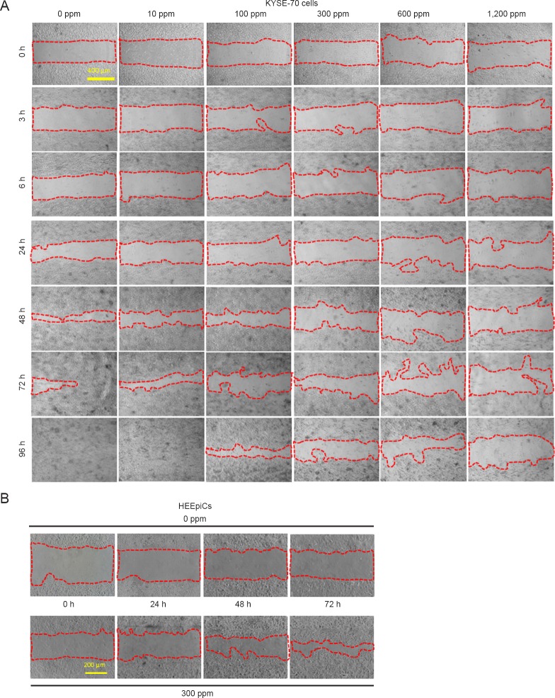
Figure 3.
Cell migration is reduced by H2-silica administration in KYSE-70 and HEEpiCs (× 40).
Note: (A) Phase contrast images of the wound-healing assay in KYSE-70 cells at 0, 3, 6, 24, 48, 72, and 96 h after wound scratching. (B) Phase contrast images of HEEpiCs at 0, 24, 48, and 72 h after chamber removal in a culture-insert migration assay. Bars: 400 μm in A, 200 μm in B. HEEpiCs: Human esophageal epithelial cells; h: hour(s); H2-silica: hydrogen-occluding-silica.

