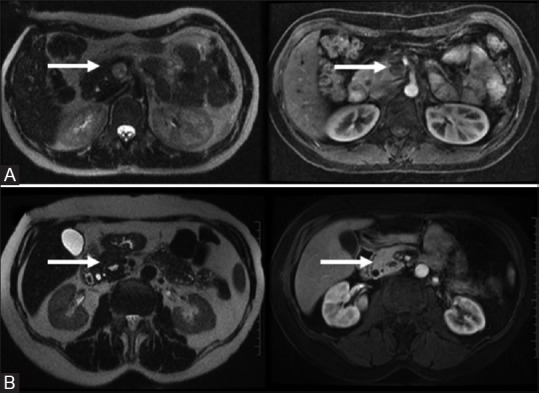
Figure 14(A and B).

Plexiform Neurofibroma: complex T2 bright “trilobar” appearance (A, top row). Solitary Fibrous Tumors: T1/T2 dark mildly enhancing wall with a central cystic component (B, bottom row)

Plexiform Neurofibroma: complex T2 bright “trilobar” appearance (A, top row). Solitary Fibrous Tumors: T1/T2 dark mildly enhancing wall with a central cystic component (B, bottom row)