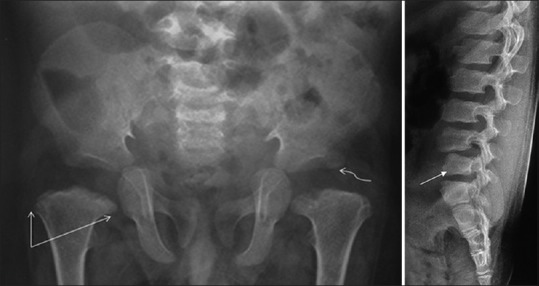
Figure 17(A and B).

SEDC with pelvic radiograph (A) demonstrating delayed ossification of bilateral femoral heads and horizontal configuration of acetabular roofs (squiggly arrow). Also note bilateral femoral metaphyseal flaring (straight arrows) and coxa vara. Radiograph of lumbosacral spine (B) in the same patient depicts anisospondyly (L4 vertebral body larger than L5 vertebral body) (arrow)
