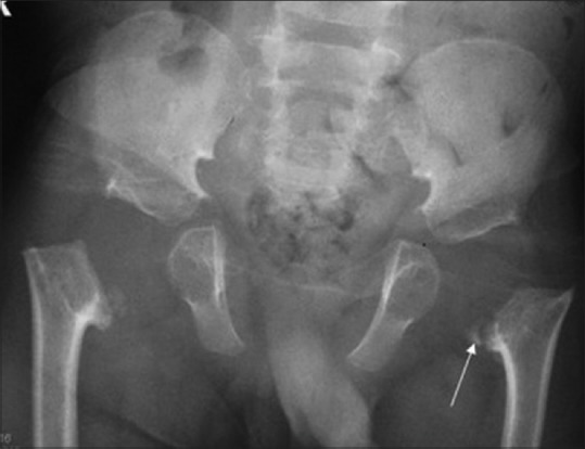
Figure 18.

SEMD: Pelvic radiograph reveals imaging findings similar to SEDC, with metaphyseal flaring and coxa vara – note the prominent metaphyseal irregularity (arrow)

SEMD: Pelvic radiograph reveals imaging findings similar to SEDC, with metaphyseal flaring and coxa vara – note the prominent metaphyseal irregularity (arrow)