
Figure 20.
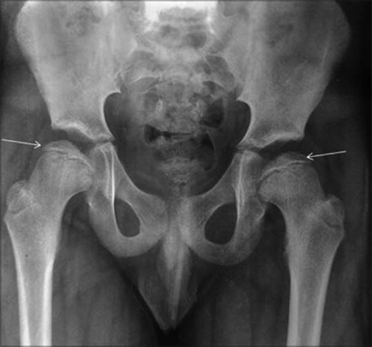
Small capital femoral epiphyses in multiple epiphyseal dysplasia. Radiograph of the pelvis shows small and irregular bilateral femoral head (marked); akin to bilateral Perthe's disease. Radiograph of the knee (not shown here) showed irregular epiphyses around the knee joint, and a tibiotalar slant was apparent in ankle radiograph