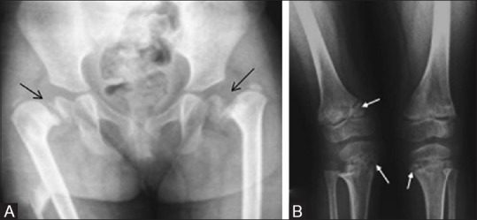
Figure 24(A and B).

Spondylometaphyseal dysplasia Sutcliffe type – (A) Pelvis with hip joint anteroposterior radiograph reveals bilateral coxa vara and slipped capital femoral epiphyses (B) bilateral knee anteroposterior radiograph reveals metaphyseal fraying with corner fractures (arrows)
