
Figure 3(A and B).
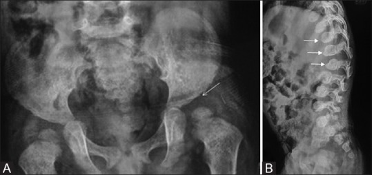
(A and B) Pelvis radiograph in mucopolysaccharidosis type I (A) reveals steep sloping of bilateral acetabular roofs (marked), which are irregular. Lumbosacral spine radiograph lateral view (B) of the same patient reveals localized angular kyphosis at the dorsolumbar junction and beak-like projection (arrows) from the inferior aspect of visualized thoracolumbar vertebral bodies (inferior beaking)