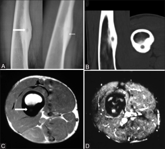
Figure 1(A-D).

Anteroposterior and lateral radiographs (A) show a radiolucent nidus (thick arrow) amid an area of fusiform cortical thickening in the diaphysis of right femur (thin arrow) better seen on sagittal and axial CT images (B). Axial MRI shows a localized T1 dark periosteal bone formation (C) with central nidal enhancement on post contrast images (D)
