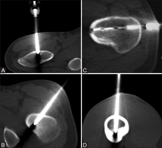
Figure 2(A-D).

Procedural axial CT images (A-D) show that a direct vertical approach (A), an oblique approach (B) to avoid the neurovascular bundle, lateral approach through opposite cortex (C) to avoid needle skidding and an anterior approach (D) for lesions in posterior cortex to obviate prone position could be used
