Figure 3(A and B).

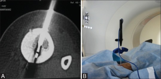
Axial CT image (A) depicting RFA probe (block arrow) across the nidus with the outer cannula of access needle withdrawn approximately 1 cm from the tip. Configuration of the RF probe and outer access needle (B) in the CT gantry

Axial CT image (A) depicting RFA probe (block arrow) across the nidus with the outer cannula of access needle withdrawn approximately 1 cm from the tip. Configuration of the RF probe and outer access needle (B) in the CT gantry