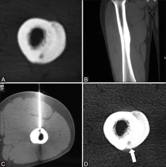
Figure 5(A-D).

Unenhanced axial (A) and sagittal (B) CT images show a small nidus in the posterior cortex of femoral diaphyses with extensive periosteal new bone formation. Procedural CT (C) shows the tip of the biopsy needle in the nidus with patient in prone position (exception). Follow up axial (D) CT images show no change in nidus size (thick arrow) or periosteal reaction
