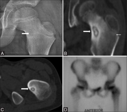
Figure 6(A-D).

AP radiograph of left hip joint (A) shows a faint radiolucent focus (thick arrow) in the neck of the left femur with no significant cortical thickening. CT coronal MPR (B) and axial (C) images show central nidal calcification with minimal reactive periosteal bone formation. Bone scan shows intense radiotracer uptake in the corresponding location (D)
