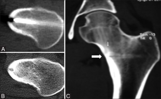
Figure 7(A-C).

Procedural CT (A) shows the tip of the biopsy needle in the nidus. Follow-up axial (B) CT images show no significant change in lesion. Because patient continued to have pain, retrospective analysis of 3D MPR coronal images (C) revealed that the biopsy track (thick arrow) is running a little inferior to the nidus (thin arrow)
