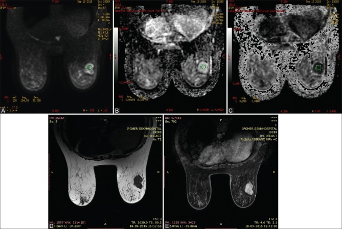
Figure 3(A-E).
37 year old woman with hyalinised fibroadenoma in right breast. Axial Diffusion-weighted imaging at b800 demonstrates high signal intensity in the right breast (A), high signal intensity on the corresponding ADC map (B) and low signal intensity in corresponding exponential ADC image (C), consistent with no diffusion restriction. The ADC and EXPONENTIAL ADC values were 0.00169mm2/s and 0.259 respectively. Axial T2 WI (D) shows a well defined hypointense lesion in the right breast. The lesion shows homogenous enhancement on post contrast study (E)

