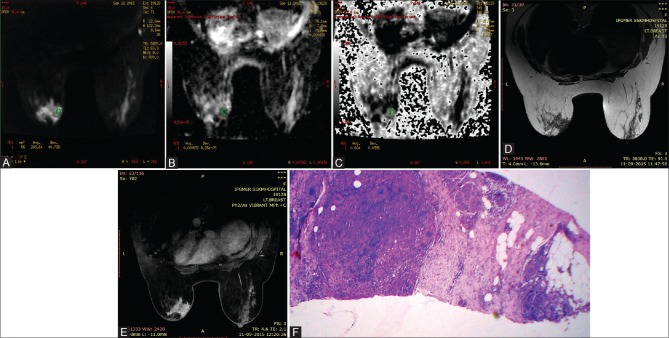
Figure 7(A-F).
49 year women with idiopathic granulomatous mastitis of left breast. Axial DWI at b800s/mm2 shows areas of hyperintensity within the mass (A) with corresponding areas of hypointensity in ADC image (B) and hyperintensity in EXPONENTIAL ADC image (C), consistent with diffusion restriction. ADC and EXPONENTIAL ADC values were 0.000632 mm2/s and 0.604 respectively. Lesion is hypointense on axial T2WI (D) & shows heterogenous enhancement in post contrast study (E) (H&E 40×10): Microphotograph shows presence of collection of epithelioid cells in fibrocollagenous stroma (F)

